Cancerous Cherry Angioma on Breast: What You Need to Know
Authored by


Skin health is crucial for overall well-being and physical comfort; abnormal growth or discoloration cannot be overlooked. Known scientifically as Campbell de Morgan spots or simply Cherry Angiomas, they warrant attention due to their potential link with malignant transformations.
Skin conditions have various complex presentations, and understanding each condition can be challenging at times, especially when dealing with a potentially cancerous one. One particular concern for many is cherry angioma on the breast, an area that naturally arouses fear due to high instances of malignancy. Although typically benign, these small red bumps occasionally indicate a more severe underlying issue, such as malignancies.
An essential assessment includes understanding what these lesions represent, how they emerge, and progress if left unchecked. Furthermore, actionable insights into effective identification techniques along with comprehensive treatment strategies might significantly alleviate concerns regarding cherry angiomas on the breast.

Cherry Angioma denotes benign skin growth. Characteristically small in size and bright red or cherry-like in appearance—hence the name—these bumps often appear on various body parts. At first sight, they are perceived merely as a cosmetic inconvenience rather than a health concern.
However, it's crucial not to overlook changes in one's physical well-being, especially when they involve potential ambiguity between harmlessness and malignancy, such as Cherry Angiomas.
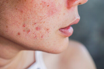
In terms of their visual representation, Cherry angiomas are typically raised from the skin surface with rounded edges and a smooth texture. Shiny papules ranging from minuscule 1 mm to 10 mm in diameter might be indicative signs.
Commonly noticed color variations span light pinkish hues, gradually intensifying into deep cherry red tones over time, sometimes even purplish undertones. There are occurrences documented showing them flat initially, then evolving towards raised formations later during life cycles.
Although predominantly prevalent among aging adults (40 years onward), instances come up among younger groups. Overall, females represent higher susceptibility compared to males—particularly those who have gone through pregnancies—due once again to largely hormonal factors at play here.
Understanding the causative factors behind cherry angiomas remains a complex task. Experts in dermatology continue to explore this still-murky science, yet some plausible explanations do exist.
One prominent theory involves aging as a significant contributor to their formation. Age-related changes in the body's vascular system give rise to these red spots predominantly among adults above 40; nevertheless, occurrences aren't limited strictly within old-age boundaries and occasionally spill over to younger demographics.
Certain research suggests potential genetic predisposition playing an influential role here - morphology similarities noted extensively across family generations back this claim convincingly.
Hormonal fluctuations such as those witnessed during pregnancy or menopause have been linked with elevated incidences, especially amongst women – suggesting hormones might trigger more widespread growths.
Exposure to certain chemicals (bromides) and climatic elements (sun damage due to UV rays) are other listed triggers attributed loosely, though not entirely supported by strong empirical evidence yet; hence, further scientific investigation into these aspects will eventually lead to a clearer understanding.
A crucial aspect demanding attention revolves around breast-specific instances of cherry angioma being perceived differently than when they emerge elsewhere on the body - primarily because breasts remain susceptible areas prone to significantly higher risk concerning malignancies or cancerous transformations compared to relatively lower-risk bodily regions historically.
Breasts naturally display extensive glandular structures interspersed inside adipose tissue. They undergo constant change cycles throughout different life stages, heavily influenced by intrinsic and extrinsic hormonal signals alike. These cycles sometimes result in irregular cell proliferation, translating outwardly visible deformities, including benign lesions like Cherry Angiomas, amidst often normal physiological responses.
Cancerous cherry angiomas are rare malignancies that stem from otherwise harmless skin lesions known as Campbell de Morgan spots or, simply, Cherry Angioma. Typically, these noncancerous crimson papules pose minimal health risks; however, any significant alterations in their structures might indicate a malignant transformation.
Differentiating between cancerous and benign formations revolves around changes in size, coloration (darkening), pattern irregularities, and expediting growth speed—combined, these clues often signal something more menacing beneath.
While initial attribution to mere cosmetic inconvenience is widespread among individuals discovering them, there are instances where symptoms start evolving beyond merely skin-deep concerns.
The presentation of such potentially dangerous variants extends towards causing itching sensations at times, even bleeding, if disturbed excessively— heightened sensitivity & discomfort correspond closely with potential malignancy.
Removal of Cherry Angiomas, particularly those located on the breast, involves several surgical procedures. The selected method often depends upon each case's specifications: the size and number of lesions, their exact location, and personal health history. Dermatologists consider all these factors.
Electrocauterization involves a process where an electric current is utilized to heat a tiny probe. The probe subsequently burns off skin anomalies while simultaneously minimizing blood loss owing to its nature of converging heat with precision, hence ensuring minimal damage to surrounding tissues.
Cryotherapy involves extreme cold delivered via liquid nitrogen onto the affected area, causing it to harden and then fall off naturally over time. It is commonly used for various dermatological conditions, including warts and moles.

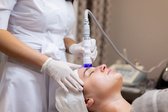
Laser therapy represents yet another viable option. Highly focused beams target the lesion's roots, thereby annihilating them without affecting surrounding healthy cells; the advantage is virtually painless compared to traditional surgeries, alongside lesser chances of recurrent issues in later stages due to the widespread success rate worldwide.
Each removal process carries inherent benefits but also potential downsides, like temporary discoloration or slight scarring after the treatment phase, which makes it imperative that patients consult experienced medical professionals before making any definitive decision concerning their well-being.
Immediate medical attention becomes necessary when any changes in an individual's appearance of cherry angiomas are observed. These alterations might include rapid growth, bleeding, itching, or a modification in the color or shape of these skin bumps.
Concern intensifies particularly if they occur on breast tissue as this area is intrinsically linked with higher cancer risks; hence, it becomes vital to arrange for a consultation immediately upon noticing such variations.
Inner wisdom says that prevention reigns supreme over cure any day. Hence, keeping watchful eyes open for early signs aids in timely intervention, eventually ensuring better health prospects.
The British Association Of Dermatologists (BAD) recommends patients regularly monitor all developments throughout body surface areas but emphasizes extra vigilance needed around breasts due to inherent malignancy tendencies occurring relatively frequently here compared to other parts.
Therefore, consulting healthcare professionals at the earliest not only supports correct diagnosis but also paves the way for devising suitable treatment plans aimed at mitigating potential complications while eradicating worries regarding possible transition into severe conditions like cancers, ultimately thus aligning oneself with a healthier life path ahead confidently.
Should I worry about cherry angiomas?
While cherry angiomas are generally harmless, any sudden changes in their appearance require medical attention.
How do you get rid of cherry angioma in the breast?
Removal of cherry angiomas often involves pulsed dye laser therapy, cryotherapy, or electrocautery. The choice depends on the patient's health and personal preference.
What skin cancer looks like a cherry angioma?
Certain skin cancers, particularly basal cell carcinoma (BCC), can mimic benign cherry angiomas. BCC often presents as shiny bumps similar to raised cherry angiomas. The resemblance between these conditions necessitates immediate medical attention for any alterations or new lesions similar to a Cherry Angioma.
When should I get cherry angiomas checked?
Immediate medical consultation is necessary if a cherry angioma changes in size, shape, or color, especially when located on the breast. This proactive approach allows for timely detection and treatment of potential underlying issues.


Plus get the inside scoop on our latest content and updates in our monthly newsletter.